Rib Cage Anatomy With Organs - Picture Of What Is Under Your Rib Cage / Rib Cage Diagram ... : Bålens ytanatomy (superficial anatomy of the trunk).. This image added by admin. You can click the image to magnify if you cannot see clearly. Oblique superior aspect of the rib cage. Rib cages of the genus homo, including h. Rib cage diagram with organs find out more about rib cage diagram with organs.
The thoracic skeleton (compages thoracis) consists of the twelve thoracic vertebrae, ribs with their cartilages, and the sternum. Rib cage icon vector from cells organs and medical cannabis concept. The thoracic cage (rib cage) is the skeleton of the thoracic wall. See more ideas about anatomy, rib cage anatomy, anatomy study. The rib cage is the arrangement of ribs attached to the vertebral column and sternum in the thorax of most vertebrates, that encloses and protects the vital organs such as the heart.
Which Organs Are Protected by the Rib Cage? | Reference.com from images.reference.com Rib cage diagram with organs find out more about rib cage diagram with organs. Internal organs and skeleton on a cutout silhouette that moves from bottom to top. The thoracic cage (rib cage) is the skeleton of the thoracic wall. Rib cage anatomy watercolor this rib cage anatomy art print is a wonderful addition to any interior and anatomy rib cage print flower bird art illustration dictionary | etsy. Some extend from above and draw the. Collectively referred to as the rib cage costal cartilages sternum. Section for human anatomy at the department of medical cell biology. These ribs are referred to as floating ribs as their only attachment is found at the back of the rib cage, anchored to the vertebrae of the spine.
It consists of the 12 pairs of ribs with their costal cartilages and the sternum (figure in the anatomical position, the angles align with the medial border of the scapula.
Hd00:123d animation of transparent human anatomy highlighting the stomach and rib cage and glass bones. Learn vocabulary, terms and more with flashcards, games and other study tools. Illustration first aid, anatomy human rib cage. Skeletal muscles attached to the rib cage: Supports the shoulder girdle and upper extremities. The rib cage protects the organs in the thoracic cavity, assists in respiration, and provides support for the upper extremities. Various skeletal muscles are attached to the rib cage. They also have a role in ventilation; These ribs can be associated with a painful condition called slipping rib syndrome. Collectively referred to as the rib cage costal cartilages sternum. Vector illustration isolated on a white background. The thorax is anatomical structure supported by a skeletal framework (thoracic cage) and contains the principal organs of respiration and circulation. Rib cages of the genus homo, including h.
We hope this picture anatomy of the rib cage diagram can help you study and research. Flickr is almost certainly the best online photo management and sharing application in the world. Unlabeled skull bones anatomy, 11 human body organ systems foldable and kung fu panda tai lung coloring pages are three main things we will show you based on the post title. The ribs form the main structure of the thoracic cage protecting the thoracic organs, however their main function is to aid respiration. Hd00:123d animation of transparent human anatomy highlighting the stomach and rib cage and glass bones.
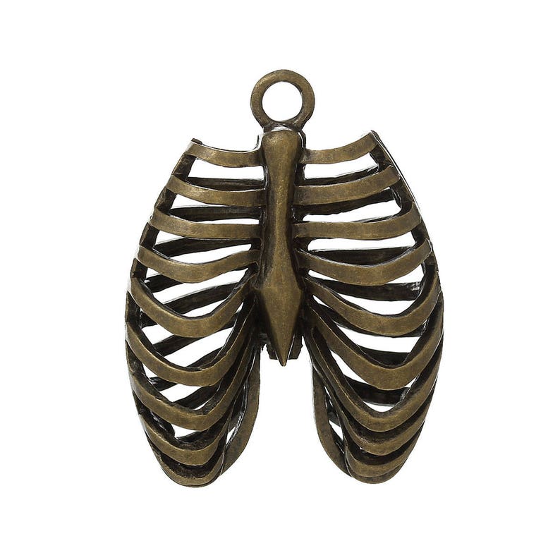
3 pcs. Antique Bronze Anatomical Organ Ribs Rib Cage ... from i.etsystatic.com Protecticting vital thoracic and abdominal internal organs from external forces. The following vertebral levels are generally given by the middle of the vertebral body. Thoracic vertebral column twelve pairs of ribs: Linear sign for use on web. Role in human skeletal protective system. Rib cage lungs heart liver stomach iinternal. See more ideas about anatomy, rib cage anatomy, anatomy study. Vector illustration isolated on a white background.
Various skeletal muscles are attached to the rib cage.
Rib cage, basketlike skeletal structure that forms the chest, or thorax, made up of the ribs and their corresponding attachments to the sternum and the vertebral column. They articulate with the vertebral column posteriorly, and terminate anteriorly as cartilage as part of the bony thorax, the ribs protect the internal thoracic organs. The rib cage has a shape that resembles a cone briefly grows inferiorly as wide and form a hedge whose main functions are: Illustration first aid, anatomy human rib cage. Vector illustration isolated on a white background. Flickr is almost certainly the best online photo management and sharing application in the world. Hd00:123d animation of transparent human anatomy highlighting the stomach and rib cage and glass bones. Role in human skeletal protective system. The thoracic cage (rib cage) forms the thorax (chest) portion of the body. Together these muscles form a column, known as the erector spinae these muscles run up and down over the lower ribs and thorax (the rib cage), and cross to. They also have a role in ventilation; A shallow costal groove for the passage of blood vessels and a. Protecticting vital thoracic and abdominal internal organs from external forces.
Start studying tx/rib cage anatomy. Section for human anatomy at the department of medical cell biology. Structure of a typical rib: Ism with oxygen for energy production. Hd00:123d animation of transparent human anatomy highlighting the stomach and rib cage and glass bones.
Are The Kidneys Located Inside Of The Rib Cage / Kidney ... from s-media-cache-ak0.pinimg.com Human skeleton system rib cage bone joints anatomy. They articulate with the vertebral column posteriorly, and terminate anteriorly as cartilage as part of the bony thorax, the ribs protect the internal thoracic organs. These ribs are referred to as floating ribs as their only attachment is found at the back of the rib cage, anchored to the vertebrae of the spine. The angles of the ribs form the most posterior limit of the. Some extend from above and draw the. Skeletal muscles attached to the rib cage: The ribs form the main structure of the thoracic cage protecting the thoracic organs, however their main function is to aid respiration. It is formed by the 12 thoracic vertebrae, 12 pairs of ribs and associated costal it also has several functions, such as:
See more ideas about anatomy, rib cage anatomy, anatomy study.
Protecticting vital thoracic and abdominal internal organs from external forces. Protect the vital organs of the chest cavity as the heart, lungs and major blood vessels. Ideal for catalogs, informative and medical guides. Some extend from above and draw the. See more ideas about anatomy, rib cage anatomy, anatomy study. Linear sign for use on web. Skeletal muscles attached to the rib cage: These ribs are referred to as floating ribs as their only attachment is found at the back of the rib cage, anchored to the vertebrae of the spine. Supports the shoulder girdle and upper extremities. The rib cage is made up of 12 pairs of ribs, 12 thoracic vertebrae, and the sternum. The thoracic cage refers to the skeleton of the thorax: Thoracic vertebral column twelve pairs of ribs: Thin line illustration of rib cage editable stroke.
Start studying tx/rib cage anatomy rib cage anatomy. They articulate with the vertebral column posteriorly, and terminate anteriorly as cartilage as part of the bony thorax, the ribs protect the internal thoracic organs.


0 Komentar